Imaging mass spectrometry is a revolutionary new technology.
The instrument is a combination of an optical microscope which allows the observation of high-resolution morphological images, with a mass spectrometer which identifies and visualizes the distribution of specific molecules.
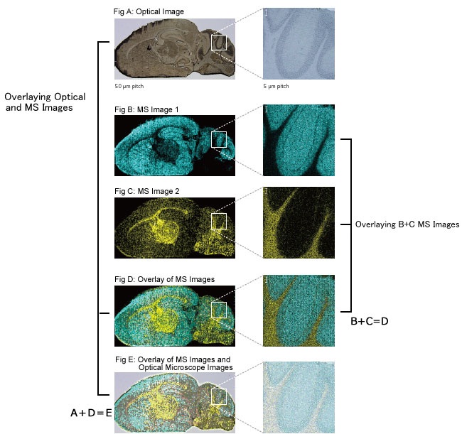
Superimposing the two images obtained based on these very different principles,has created a significant new research tool, the imaging mass microscope. The accurate and high resolution mass images from the iMScope will drive your research to the next level.
At long last, we have entered the age of imaging mass spectrometry.
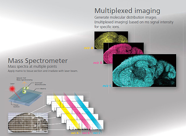
Imaging mass spectrometry directly detects both natural and synthetic molecules in tissue sections and measures mass spectra while preserving location information associated with the tissue section.
Two-dimensional distributions of specific molecules are then visualized by combining the position information of each mass spectrum and the signal intensity for specific ions in the mass spectrum. (MS imaging).
The iMScope TRIO imaging mass microscope is an instrument designed specifically for imaging mass spectrometry. It represents a hybrid type microscope that combines both an optical microscope and a mass spectrometer. iMScope TRIO now enables the direct identification of various substances in tissue samples, expanding potential research opportunities in a wide variety of fields.
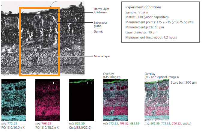
The high resolution imaging offered by optical microscopes is essential not only for pharmacokinetic analysis, but also for toxicity testing and toxicity mechanism analysis. Analysis of retina and skin requires high spatial resolution imaging.
In this experiment, the fat in rat skin was visualized. High spatial resolution imaging clearly showed localization of ceramide in the horny layer and phosphatidylcholine (PC) in the sebaceous gland.
In pharmacokinetic analysis, the iMScope TRIO provides simultaneous mapping of the distribution of unchanged and metabolized drugs in a single measurement without any labeling. The laser diameter used during mass spectrometry imaging with the iMScope TRIO ranges continuously from 5 to 200 µm, offering low to high spatial resolution. This helps ensure that analyzes are performed as efficiently as possible.
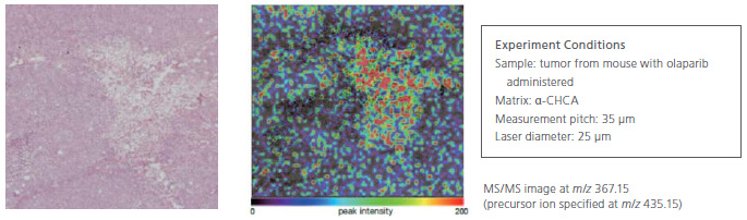
Using a mouse administered with olaparib, the pharmacokinetic status of the tumor was visualized. In this way, imaging mass spectrometry is being utilized even in the clinical trial phase of drug discovery, rather than only during basic research.
Overlaying MS images and optical microscope morphological images reveals the difference between the amounts of
specific molecules in each minute organ and can relate molecular distribution to the biological functions of an organelle and morphological changes.
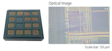
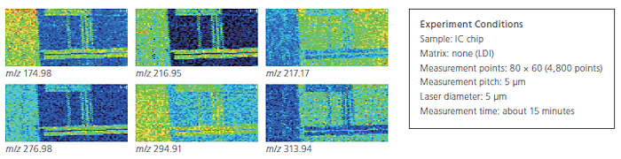
In industrial fields, various surface inspection technologies are widely used to ensure stable production of high quality products. Imaging mass spectrometry is capable of analyzing various contaminants that cannot be detected by conventional surface inspection methods, and it is possible to gain new knowledge for higher quality production.
An example using IC chip is shown. A wide variety of optical images and mass spectrometry imaging technologies are used to support cutting-edge research.
 Loading...
Think big see beyond... Ant Teknik is is offering sales, service and spare part supply; validation, application and training as well your turn-key laboratory project services for research and QC laboratories...
What is m/z? what does S/N ratio mean? What does is included in OQ? Explore answers to the most common questions we get from analysts…
Tips from our experts on chromatography, spectroscopy and sample preparation that you can benefit from in analytical laboratory...