Thermo Fisher Scientific

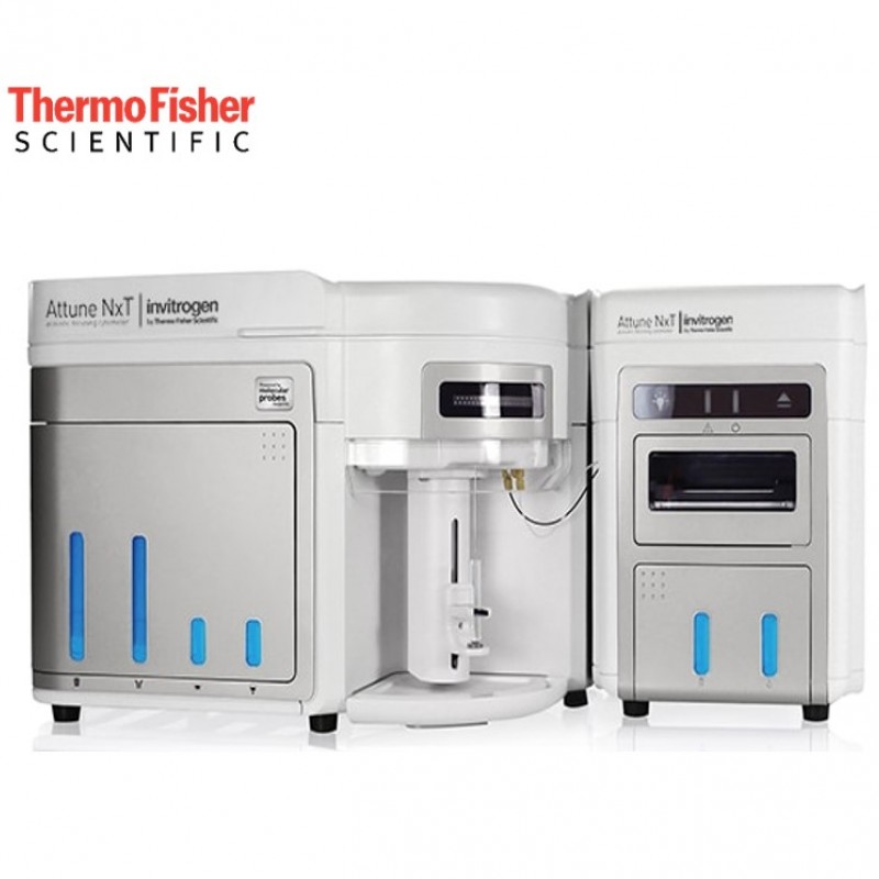
Invitrogen™ Attune™ NxT Flow Cytometer

The Invitrogen™ Attune™ NxT Flow Cytometer is an advanced cell analyzer where acoustic focusing fluids are key to both high sensitivity and high efficiency.

Attune™  NxT

The Attune NxT Flow Cytometer shares common features with the Attune CytPix model.
  • Acoustic focusing for precision
  • Fast, accurate purchase
  • Rare event detection
  • Designed for flexibility
  • New optical design
  • Clog resistant technology
  • Powerful, intuitive software
  • Biosafety header compatible

Flow Cytometry Applications

Almost any standard flow cytometry application can benefit from the high sensitivity and throughput of the Attune NxT Flow Cytometer. See our Sample Data page for examples spanning infectious disease research, fluorescent proteins, CRISPR gene editing, immuno-oncology research, microbiology, plant ploidy, platelets, stem cells, and T cells.

One classic flow cytometry application is immunophenotyping. Flow cytometry is the method of choice for identifying cells within complex heterogeneous populations, as it allows for multiparameter analysis of thousands to millions of cells in a short time. Strong signal separation in the Attune NxT Flow Cytometer shows excellent separation of cell populations into subsets for immunophenotyping. A wide range of reagent choices—as well as the system’s automated compensation module, four spatially separated lasers, and 14 color choices—help to simplify multicolor panel design.

Thirteen color immunophenotyping using Attune NxT


Gating strategy for 13-color immunophenotyping analysis of stained human whole blood using a stain/lyse protocol. Human whole blood cells
were stained as described in the application note and acquired and analyzed on the Attune NxT Flow Cytometer. (A) Dead cells were excluded
from the analysis by gating on live (propidium iodide–) cells in a dot plot. (B) CD45+ gating was used to select the leukocyte population from the
lysed whole blood. (C) Lymphocytes and monocytes were identified based on forward and side scatter profiles. (D) Monocytes are found above
lymphocytes on the scatter plot and express both CD14 and CD33. (F) Within the lymphocyte gate, immune cells can be subdivided based on
their expression of CD3 (T cells), CD19 (B cells), or neither (NK cells). (E) B cells can be further characterized by HLA-DR and CD45RA expression.
(G) T cells can be further subdivided into CD4+ (T helper cells) and CD8+ (cytotoxic T cells) subpopulations, while (J) regulatory T cells express
CD4 and CD25. (H, K) CD62L identifies naive (TN) CD4 and CD8 T cells, while HLA-DR is expressed by activated T cells (TA). (I) Finally, NK
cells lack B cell and T cell markers (CD19–CD3–) and express CD56.

For more information please click.

Recommend Me a System

Please Recommend Me a Column