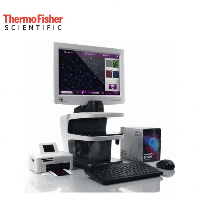
The FLoid® Cell Imaging Station is a fully integrated devise seamlessly combining high-quality optics, detection in three fluorescence channels as well as transmitted light, a widescreen LCD monitor, camera, image acquisition/processing software, and reagent information and protocols. It features a fixed, high-quality 20x plan fluorite objective, with additional magnification obtainable using the digital zoom. Fluorescence is captured in the three most commonly used channels (DAPI, FITC, and Texas Red filters). Transmitted light images automatically show cells in excellent contrast through a relief phase design, without loss of light. With its innovative light shield, ambient light is blocked so the FLoid® Cell Imaging Station can be placed wherever you need it most - in the tissue culture room, among other cell-based assay instruments, or on your lab bench. FLoid® Cell Imaging Station lets you bring imaging out of the darkroom.
 |
 |
|
Induced human pluripotent stem cells |
Fixed Indian Muntjac cells |
 |
 |
|
Autofluorescent fly wing |
Live phagocytosis in MMM cells |
The FLoid® Cell Imaging Station uses an open-stage design providing maximum vessel flexibility for slides, multi-well culture dishes, or even T75 flasks. A focus assist scale makes it easy to find the focus plane. Once samples are in focus, you can use the mouse to change light intensity and switch colors; there is also a scale bar that changes as the digital zoom level is changed, making it easy to estimate and compare sample sizes. The absolute magnification span is from 460X up to 1,840X using the digital zoom. Images can be printed directly using the optional printer, or saved for viewing in one of the four formats (JPEG, TIFF, BMP, or PNG) at a later time or with other software. With printer you can generate sticker-backed prints for immediate notebook mounting. The FLoid® Printer is not included with the FLoid® Cell Imaging Station and must be purchased separately.
Designed to be one of the simplest, fastest, and easiest cell imaging systems in the world, the EVOS FLoid system captures high-quality, 3-color fluorescent cell images right at your benchtop within a minute. Usage is so intuitive, even novice users can collect data in just a few mouse clicks. The EVOS FLoid system is ideal for cell culture applications requiring fluorescence, or for just giving you a quick view of your cells.
| Light Source | Excitation (nm) | Emission(nm) | Compatible Dyes |
| Blue | 390/40 | 446/33 | DAPI, Hoechst stain, NucBlue, Alexa Fluor 350, BFP |
| Green | 482/18 | 532/59 | GFP, Alexa Fluor 488, Calcein, SYBR Green, FITC, Fluo-4, pHrodo Green |
| Red | 586/15 | 646/68 | RFP, Alexa Fluor 546/555/568, PE, Cy3, MitoTracker Orange, pHrodo Red, Rhodamine Red, DsRed |
By incorporating the Molecular Probes® reagent selection guide, the FLoid® Cell Imaging Station simplifies fluorescent dye selection and cell staining. You can easily access detailed information on over 160 reagents in 20 application areas directly from the user interface. Each reagent has a quick reference protocol with icons and concise text for ease of use. These reagents have been validated for spectral compatibility with FLoid®, so you can obtain optimal images.
Specifications |
|
| Objective | 20X plan fluorite |
| Numerical aperture (N/A) | 0.45 |
| Working distance (WD) | 5.9 mm (defined as the tip of the objective to the bottom of a 1.2-mm thick slide) |
| Overall magnification | 460X (optical)-1840X (with digital zoom) |
| Display resolution | 1366 x 768 pixel |
| Image resolution | 1296 x 964 pixel |
| Contrast methods | Fluorescence and transmitted light (relief phase) |
| Color channels | 4 channels (relief phase, blue fluorescence, green fluorescence, red fluorescence) |
| Illumination | LED (50,000 hour life), adjustable intensity |
| Excitation | Blue channel: 390/40 nm; Green channel: 482/18 nm; Red channel: 586/15 nm |
| Emission | Blue channel: 446/33 nm; Green channel: 532/59 nm; Red channel: 646/68 nm |
| Stage to condenser | 60 nm |
| Camera | Sony 1.3MP 1/3” ICX445 EXview HAD CCD |
| Instrument type | Benchtop device for imaging cells |
| Dimensions | 15.9” (W) x 21.1” (H) x 13.9” (D) 40.4 cm (W) x 53.6 cm (H) x 35.3 cm (D) |
| Weight | 26 lbs (11.8 kg) |
| Operating power | 100-240 VAC |
| Frequency | 50-60 Hz |
| Electrical input | 5 VDC, 4.15 A |
| Working temperature | 4-32ºC |
| Working humidity | <%90 (non-condensing) |
| Outlet port | 4 USB |
| Stage | Mechanical XY glide stage that can move within a 5 mm diameter circle |
| LCD Monitor | 15-inch color; display resolution 1366 x 768 pixels; Image resolution 1296 x 964 pixels; adjustable tilt |
| Software | FLoid Cell Imaging Station Software |
| Captured images | 16-bit monochochrome TIFF, PNG, BMP and JPG; 1296 x 964 pixels |
| Supplied USB drive | 2 Gigabyte |
The key feature shared by all EVOS cell imaging systems is exceptional image quality. Capture clear, bright, broadcast-quality images and videos to help tell the story of your data in just a few clicks.
 |
 |
| HeLa cell large field vs. resolved EVOS M7000, 60x objective, Celleste Image Analysis Software. Stains: NucBlue (core), Phalloidin. |
11-slice image Z-stack of honeybee claw EVOS M5000 in color mode, 10x Apo objective |
 |
 |
| Induced human pluripotent stem cells EVOS Floid. Stains: Lin28A (green), Tubulin (red), Hoechst (blue). |
Mouse melanocytes EVOS XL Core, 20x. |
 |
 |
| Z-stack movie “Walk through” the Z-dimension of an object in 0.15-µm layers. |
Wound healing EVOS M7000, 10x, Stage Incubator over 21 hours. Stains: DsRed-cadherin-4, GFP. |
Depending on the model, EVOS cell imaging systems are a high-sensitivity monochrome camera ideal for fluorescent imaging; high sensitivity color camera ideal for colorimetric imaging; or both monochrome and color cameras for maximum flexibility.
A choice of 30+ high-performance objective lenses ranging from 1.25–100x allow you to adjust the optics for imaging type, sample type and vessel. Image acquisition is fast and with automated multi-well scanning, the M7000 can scan a 96-well plate in less than 5 minutes across 3 fluorescent channels. EVOS cell imaging systems make it easy to capture broadcast quality images in seconds.
All EVOS microscopes come with onboard software that allows you to view, capture, and process images quickly and easily. You can:
On the M7000 and M5000, the onboard software offers pinpoint operational control and powerful image processing tools such as cell counting, confluence, Z-stacking, image tiling and stitching, time-lapse movies, and RGB synthesis. These tools operate intuitively with minimal training, aided by onboard SmartStart guides. Autofocus and other automated tools save time and effort. The M7000 adds advanced automation features and complete programmatic control, allowing imaging through software alone, without ever touching the instrument. Download the most recent EVOS M7000 and M5000 software updates.
With all EVOS imaging systems, a large, bright, high-definition monitor lets you view your cells outside the confines of a darkroom and facilitates safe collaboration in nearly any space. There’s no need to remove safety goggles to look through an ocular eyepiece. This minimizes difficult inter-user decontamination and facilitates both collaboration and safety.


Images from all EVOS instruments can be sorted and analyzed with the EVOS Image Analysis app in Connect, Thermo Fisher’s secure, cloud-based platform for data storage, scientific analysis apps, and peer collaboration tools. Once uploaded, you and your colleagues can collaboratively access your gallery and retrieve, analyze, and manipulate your images from any web browser around the world. You can also run other Connect applications and monitor the status and configuration of your connected instruments.

 Loading...
Think big see beyond... Ant Teknik is is offering sales, service and spare part supply; validation, application and training as well your turn-key laboratory project services for research and QC laboratories...
What is m/z? what does S/N ratio mean? What does is included in OQ? Explore answers to the most common questions we get from analysts…
Tips from our experts on chromatography, spectroscopy and sample preparation that you can benefit from in analytical laboratory...