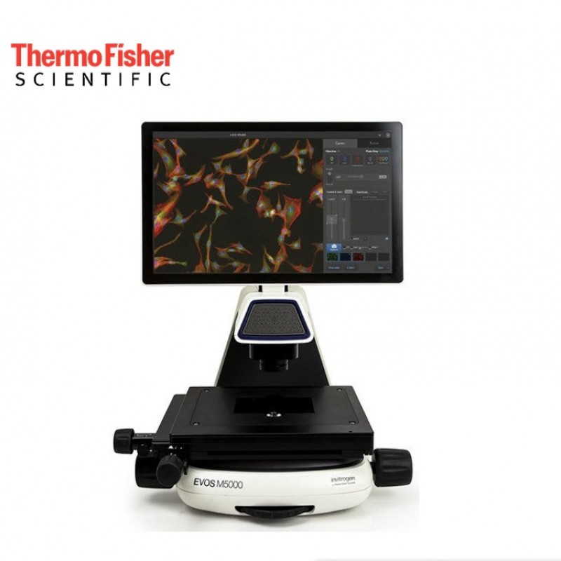
The EVOS M5000 microscope combines precision components with a unique modern design functionality that enables high-quality fluorescence and color imaging anywhere with unprecedented flexibility. It is a fully integrated imaging system that combines precision optics, an 18.5” high-resolution articulated LCD monitor, and a highly sensitive 3.2 MP monochrome CMOS camera (2048 x 1536) with 3.45-µm pixel resolution. The monochrome camera affords the best sensitivity for detection of faint fluorescence signals and allows quantitative analysis. At the same time the condenser includes a novel LED-based RGB illumination scheme that enables high-quality, low-noise color image acquisition using the same monochrome sensor, enabling rendering of true color in transmitted light, which is beneficial when imaging stained tissue samples such as H&E stained tissue sections.
At the same time, the concentrator includes a new LED-based RGB lighting scheme that enables high-quality, low-noise color images using the same monochrome sensor. It provides true color rendering in transmitted light, which is useful when viewing stained tissue samples, such as HE-stained tissue sections. |
 |
|
11-slice image Z-stack of honeybee claw |
Differentiated mouse embryonic stem cells |
 |
 |
|
HeLa cells with stained mitochondria |
Human adenocarcinoma section |
 |
 |
|
Section of rat intestine |
|
With a five-position objective turret and the ability to acquire images in four fluorescence channels, transmitted light phase-contrast, and color, the optical system can be easily configured to meet your needs. Choose from our broad range of high-quality long-working-distance and coverslip-corrected objectives for use with plastic vessels or glass sides. Available from 1.25X to 100X magnification with achromat, fluorite, or apochromat correction, together with over twenty LED light cubes, the sample imaging options are limitless. Interchangeable vessel holders accommodate most vessel types and sizes, including slides, multi-well plates, culture flasks, and Petri dishes, and afford precise control and sample alignment by the automated stage.
EVOS microscopes are designed with scientists’ workspace and workflow needs in mind. Components and controls are conveniently integrated into a single, lightweight system that fits on a benchtop and can easily be moved to classrooms, teaching labs, and conference rooms. The size and portablility of EVOS microscopes let you capture and view images when and where needed.

All EVOS imaging systems feature bright LED light sources that provide more than 50,000 hours of brilliant (yet adjustable) illumination that (unlike mercury arc lamps) remains constant with minimal degradation over time. Cost-efficient and environmentally friendly, EVOS LED light cubes are designed for precise control, minimal maintenance, and exceptional reliability.
For the M7000 and M5000, a choice of compact, easily interchangeable LED light cubes can be matched to the spectra of specific dyes or fluorescent proteins in your application. Each cube incorporates excitation filter, dichroic beam-splitter, and emission filter. Filters are hard-coated for maximum transmission efficiency.

Mercury arc lamps can lose 50% of their intensity in the first 100 hours of operation! In addition, images acquired in different sessions cannot be quantitatively compared using without complicated calibrations. Because EVOS LED light sources produce constant light intensity, users can rely on consistent illumination and can compare results quantitatively from images acquired on different days.

The LED bulbs on the EVOS systems are rated for >50,000 hours (~17 years), compared to 300 hours for a typical mercury bulb (and 1,500 hours for a metal halide bulb). That means significant savings in the overall upkeep of your instrument.

Hard-coated filter sets are more expensive but have sharper edges and significantly higher transmission efficiencies that typically result in >25% more light transmission than traditional soft-coated filters. With the EVOS system’s hard-coated filter sets, your light cubes cost less over time—and provide brighter fluorescence, higher transmission efficiencies, the ability to detect faint fluorescence signals, and better signal-to-noise ratios.
![]()
Depending on the model, EVOS cell imaging systems are a high-sensitivity monochrome camera ideal for fluorescent imaging; high sensitivity color camera ideal for colorimetric imaging; or both monochrome and color cameras for maximum flexibility.
A choice of 30+ high-performance objective lenses ranging from 1.25–100x allow you to adjust the optics for imaging type, sample type and vessel. Image acquisition is fast and with automated multi-well scanning, the M7000 can scan a 96-well plate in less than 5 minutes across 3 fluorescent channels. EVOS cell imaging systems make it easy to capture broadcast quality images in seconds.
All EVOS microscopes come with onboard software that allows you to view, capture, and process images quickly and easily. You can:
On the M7000 and M5000, the onboard software offers pinpoint operational control and powerful image processing tools such as cell counting, confluence, Z-stacking, image tiling and stitching, time-lapse movies, and RGB synthesis. These tools operate intuitively with minimal training, aided by onboard SmartStart guides. Autofocus and other automated tools save time and effort. The M7000 adds advanced automation features and complete programmatic control, allowing imaging through software alone, without ever touching the instrument. Download the most recent EVOS M7000 and M5000 software updates.

With all EVOS imaging systems, a large, bright, high-definition monitor lets you view your cells outside the confines of a darkroom and facilitates safe collaboration in nearly any space. There’s no need to remove safety goggles to look through an ocular eyepiece. This minimizes difficult inter-user decontamination and facilitates both collaboration and safety.


Images from all EVOS instruments can be sorted and analyzed with the EVOS Image Analysis app in Connect, Thermo Fisher’s secure, cloud-based platform for data storage, scientific analysis apps, and peer collaboration tools. Once uploaded, you and your colleagues can collaboratively access your gallery and retrieve, analyze, and manipulate your images from any web browser around the world. You can also run other Connect applications and monitor the status and configuration of your connected instruments.

Specifications |
|
| Camera Type | 3.2 Megapixel, monochrome, CMOS |
| Color | Assorted colors |
| Dimensions (LxWxH) | 18 in. (W) x 23 in. (H) x 18 in. (D) |
| Display | 18.5 in. articulated LCD |
| Model | EVOS M5000 |
| Product Type | Imaging System |
| Resolution | 1920 x 1080 pixel |
| Lighting Type | LED |
| Magnification Power | 1.25X to 100X |
| Objective Types | 5 position turret |
| Weight | 36 lb. |
 Loading...
Think big see beyond... Ant Teknik is is offering sales, service and spare part supply; validation, application and training as well your turn-key laboratory project services for research and QC laboratories...
What is m/z? what does S/N ratio mean? What does is included in OQ? Explore answers to the most common questions we get from analysts…
Tips from our experts on chromatography, spectroscopy and sample preparation that you can benefit from in analytical laboratory...