Thermo Fisher Scientific

Thermo Scientific™ CellInsight™ CX7 | High-Content Analysis (HCA) Platform

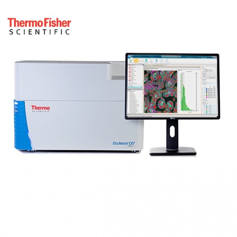
The Thermo Scientific™ CellInsight™ CX7 High Content Analysis (HCA) Platform is a fast, automated cellular imaging and analysis platform designed for quantitative microscopy.

The CellInsight CX7 HCA Platform provides an integrated way to develop and automate high-content assays using a combination of 5-color brightfield and 7-color fluorescent imaging in confocal or widefield modes.

For this particular configuration, a version with HCS Studio™ Cell Analysis Software and Store Image and Database Management Software is also available to visualize and share high content data and images.

The Thermo Scientific CellInsight CX7 High Content Scanning Platform is an LED-based platform that offers a variety of imaging modes to extract the information you need from your samples. You can choose the right modes to read your sample, both individually and on a channel basis, with the resolution and dynamic range resulting from the high performance of the optical train and precision camera. Use the entire fluorescence spectrum to optimize your assay and choose wide field or confocal optics for any channel.

Features of CellInsight CX7 HCA Platform

  • 7-channel fluorescence imaging
  • 5-channel bright field imaging
  • 7-channel confocal imaging
  • Software and laser-based autofocus for consistent scan times
  • Configured for fully automatic plate processing and scanning

Capabilities

  • Addresses hundreds of assay types with flexible software and hardware tools
  • Scan and analyze microscope slides or most SBS-standard format microplates
  • Use HCS Studio software for integrated instrument control and data analysis
  • Achieve the fastest time to results by using intelligent acQuisition (iQ) real-time analysis feedback
  • Compatible with Molecular Probes™ fluorescent detection reagents
HeLa cells imaged using the CellInsight CX7 platform
7_A549-cell
HeLa cells imaged using the CellInsight CX7 platform
HeLa cell image acquired using the Thermo Scientific CellInsight CX7 platform and stained with Invitrogen HCS NuclearMask Blue (blue), Invitrogen MitoTracker Orange (green), and Invitrogen Alexa Fluor 647 Phalloidin (red).
A549 cells imaged using the CellInsight CX7 platform
A549 cell image acquired using the Thermo Scientific CellInsight CX7 platform and stained with Invitrogen HCS CellMask Blue (blue), Invitrogen Alexa Fluor 488 Phalloidin (green), and Invitrogen Alexa Fluor 750 goat anti–mouse IgG secondary antibody (pink).
6_Human-breast-cancer-tissue
hela-488tubulin-confocal-led-60x-7600-(1)-600-dpi
Human breast cancer tissue section imaged using the CellInsight CX7 platform
Three-color brightfield image of fixed human breast cancer tissue stained with KI67-DAB and counterstained with hematoxylin and eosin and imaged with the Thermo Scientific CellInsight CX7 platform.
HeLa cells imaged using the CellInsight CX7 LED instrument
HeLa cells labeled with tubulin antibody followed by Invitrogen Alexa Fluor Plus 488 antibody and Hoechst 34580 staining. Imaging was performed on the CellInsight CX7 LED instrument using a 60x objective and confocal mode.
HeLa-650-tubulin-60x-1-LED-(2)600-(1)-600-dpi-nucleus
HeLa-mito-AF488-60X-1-LED-(1)600-(1)-600dpi
HeLa cells imaged using the CellInsight CX7 LED instrument
HeLa cells labeled with tubulin antibody followed by Invitrogen Alexa Fluor Plus 647 antibody and Hoechst 34580 staining. Imaging was performed on the CellInsight CX7 LED instrument using a 60x objective and confocal mode.
HeLa cells imaged using the CellInsight CX7 LED instrument
HeLa cells labeled with ATP synthase V antibody followed by Alexa Fluor Plus 488 antibody and Hoechst 34580 staining. Imaging was performed on the CellInsight CX7 LED instrument using a 60x objective and confocal mode.
 

Versatility and Accelerated Output with Automatic Imaging

The CellInsight family of HCS/HCA platforms offers widefield, confocal or brightfield automated imaging modes, all within a single protocol. Designed with speed in mind, real-time imaging and analysis can be run in parallel for the fastest possible turnaround time.

Publication Quality Imaging

The CellInsight family of high-content screening (HCS)/high-content analysis (HCA) platforms are designed to provide automated imaging versatility and accelerated throughput for the fastest time-to-data possible. Choose from widefield, confocal, or brightfield automated imaging modes, all within one protocol. Our instruments provide up to 12 colors to choose from, from ultraviolet through near-infrared fluorescence and multicolor brightfield imaging to enable flexible imaging paradigms. Finally, our systems are engineered with speed in mind, enabling real-time imaging and analysis conducted in parallel for the fastest time-to-data possible.   


Multicolor live-cell whole-spheroid imaging using Thermo Scientific CellInsight CX7 LZR HCS Platform in confocal mode

Onboard Real Time Analysis Software

The Thermo Scientific HCS Studio Cell Analysis Software expertly balances flexibility and ease of use to enable meaningful data analysis from single cells through populations. Parallel imaging and analysis offers true real-time results, fast. 

HCS Studio software allows backtracking of each individual cell or cellular event to minimize artifacts in the data. View the corresponding cell level cutout associated with each data point to ensure quality control of what you are actually measuring. 


Illustration showing how EurekaScan Finder software finds objects in tissue samples using 4x and 20x scanning passes

To accelerate assay development, every parameter can be evaluated for assay performance based on Z-prime statistical analysis.  This allows you to select your top-performing assays as you scale your screening campaigns. HCS Studio software also features guided workflows with user-friendly visual feedback to facilitate easy assay protocol setup, image acquisition, analysis, and scanning.

Illustration showing how EurekaScan Finder software finds spheroids and rare events using 4x and 20x scanning passes

The HCS Studio software for CellInsight CX7 LED and CellInsight CX7 LZR also feature the Thermo Scientific EurekaScan Finder software. This software significantly reduces your instrument runtime and analysis efforts by automating the detection and subsequent capture of events at higher magnification.

EurekaScan Finder software offers two different approaches based on the type of sample and user-stipulated criteria that will inform high-content screening.
 

Illustration showing the process of image import and conversion, insertion of data into database, and data/image export from database.
 

Specifications

Camera

Photometrics High-Resolution Fluorescent Camera:

  • High-resolution, thermoelectrically cooled to 0°C
  • FOV 2208 x 2208-pixel array
  • 4.54-micron pixel size
  • 75% peak quantum efficiency (QE)
Light source

LED, solid-state 7-color light engine used with provided filter sets offers the following excitation/emission capabilities (Ex/Em, in nm):

  • 386/440 (e.g., Alexa Fluor 350 dye)
  • 438/480 (e.g., Alexa Fluor 405 dye)
  • 485/521 (e.g., Alexa Fluor 488 dye)
  • 549/600 (e.g., Alexa Fluor 555 dye)
  • 560/607 (e.g., Alexa Fluor 594 dye)
  • 650/694 (e.g., Alexa Fluor 647 dye)
  • 740/810 (e.g., Alexa Fluor 750 dye)
Objectives

Standard (Olympus™ objectives):

  • 10x (0.30NA)
  • 20x (0.4NA)
  • 20x (0.7 NA) Olympus objectives
  • 40x (0.6 NA) Olympus objectives

Optional (Olympus objectives):

  • 2x (0.08NA)
  • 4x (0.16NA)
  • 40x (0.95 NA)
  • 60x (0.90 NA)
Image compability JPG, BMP, GIF, PNG, TIF, C01, DIB
Dimensions 20 in x 32 in x 18 in (50.8 cm x 81.3 cm x 45.7 cm)
Weight 68 kg (150 lb)
Data management Compatible with Thermo Scientific Store Image and Database Management Software
Software Thermo Scientific HCS Studio 4.0 Cell Analysis Software
Wattage 300 W

For more information please click.

Recommend Me a System

Please Recommend Me a Column