What is Western Blot?
Western blot is a widely used method for protein detection and identification of specific proteins in protein solution in areas such as protein biology, immunogenetics, and molecular biology.
Western blot technique combines the specificity of antibody detection with the resolution of gel electrophoresis.
Western Blot Stages
- Sample preparation
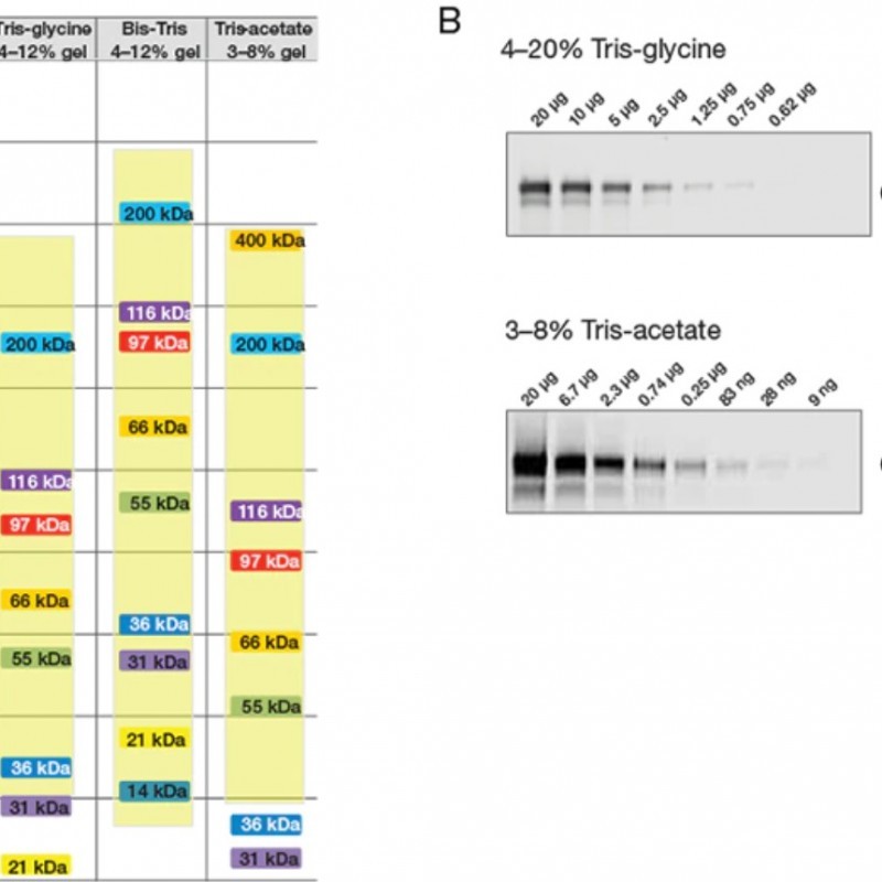
- Gel electrophoresis
- Transfer of proteins to the membrane
- Antibody incubation
- Imaging and analysis
How is Western Blot applied?
The first step in the Western blot procedure is to prepare the sample. Next, macromolecules in a sample are separated using gel electrophoresis. The separated molecules are then transferred or blotted onto a second matrix, usually a nitrocellulose or polyvinylidene difluoride (PVDF) membrane. The membrane is then blocked to prevent any non-specific binding of antibodies to the surface of the membrane.
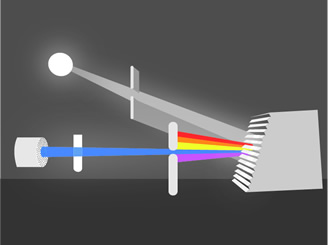
Most commonly, the transferred protein is then probed with a combination of antibodies: an antibody specific to the protein of interest (primary antibody) and another antibody specific to the host type of the primary antibody (secondary antibody). Usually, the secondary antibody complexes with an enzyme which, when combined with a suitable substrate, will produce a detectable signal. Chromogenic substrates produce a precipitate that results in visible colorimetric changes on the membrane. The most sensitive detection methods use a chemiluminescent substrate that produces light as a byproduct of the reaction with the enzyme conjugated to the antibody. Light output can be captured using film. However, digital imaging devices based on charge-coupled device (CCD) cameras are becoming popular alternatives to film for capturing the chemiluminescent signal. Alternatively, fluorescently labeled antibodies can be used that require detection using an instrument capable of capturing the fluorescent signal. Fluorescent staining is a newer technique and is growing in popularity as it provides the potential for multiplexing (detecting multiple proteins on a single stain). Whatever system is used, the intensity of the signal should be related to the abundance of antigen on the membrane.
Procedures for the detection phase of a western blot experiment vary widely. A common variation involves direct and indirect sensing. The direct detection method uses an enzyme or fluorophore-conjugated primary antibody to detect the antigen of interest on the stain. This detection method is not widely used since many researchers prefer the indirect detection method for various reasons. In the indirect detection method, an unlabeled primary antibody is first used to bind to the antigen. The primary antibody is then detected using an enzyme or fluorophore-conjugated secondary antibody. Labels (or conjugated molecules) may contain fluorescent probes such as biotin, Invitrogen Alexa Flour or DyLight fluorophores, and enzyme conjugates such as horseradish peroxidase (HRP) or alkaline phosphatase (AP). The indirect method offers many advantages over the direct method described below.
Protein Identification
Protein identification uses substrates that will react with the enzyme conjugated to the secondary antibody to form a signal to detect specific bindings on the membrane.
1-Colorimetric Identification
2- Chemiluminescent Identification
Chemiluminescence or chemiluminescence is the state of very small amount of thermal radiation and light radiation as a result of the chemical reaction taking place within the substance. In Western blot, irradiation occurs when the enzyme-specific substrate bound to the secondary antibody is placed in the medium.
3- Fluorescent Identification
In the fluorescent identification method, the secondary antibody is bound with a fluorescent maker such as flurescein, texas red, and phycoerythrin. In this method, an additional imaging system is needed to identify the fluorescent radiation.